Chondrodermatitis Nodularis Helicis (CNH): 78y F. Patient.Roy F. Sullivan, Ph.D. (posted 05/02/2001)
CASE STUDY (WARR0815)This 78 year old female patient, having a mild, bilateral SNHL, has been known to the practice for fifteen years. She presented with a chief complaint (cc) of otalgia AD and an inability to fully insert her four year old Class II (vented; 2mm internal diameter) canal hearing aid. Visual / Video Oto-Macroscopic (VOM) examination of the pinna revealed two lesions of the helix and antihelix, respectively [inline image below (29k)]. The lower, healing, abrasion was self-induced with a fingernail. The mid-anti-helical lesion, nodular, hyperaemic and painful to the touch, was diagnosed by her dermatologist as chondrodermatitis nodularis helicis (CNH). The mid-nodular scab was the site of intralesional cortisone injections. 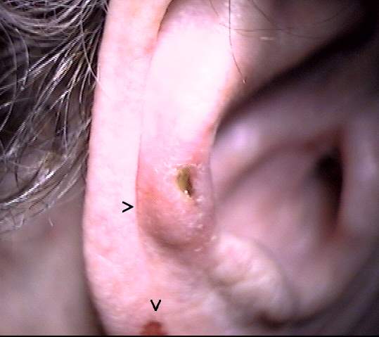 Video Oto-Endoscopic (VOE) examination of the EAC revealed an otitis externa, acute (OEA) with attendant hyperaemia, characteristic "chicken skin" papillation, canal stenosis and greenish mucoid exudate [inline image below (23k)], precluding full insertion of the canal hearing aid. The patient's dermatologist and ENT were contacted and the patient referred back for follow-up. It was recommended that hearing aid use be suspended until the EAC condition had been medically resolved. 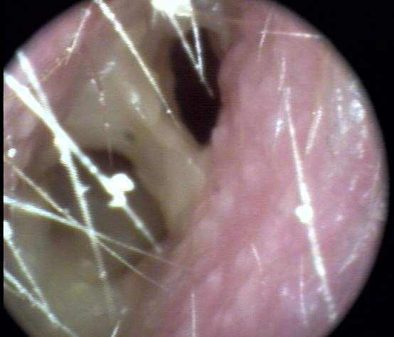 VOM and VOE images are presented after two weeks of dermatological and otological intervention. Additional intralesional injections by the dermatologist into the CNH site resulted in a significant symptomatic remission of the pinna [inline image below (20k)]. 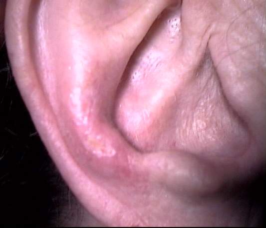 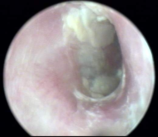 |