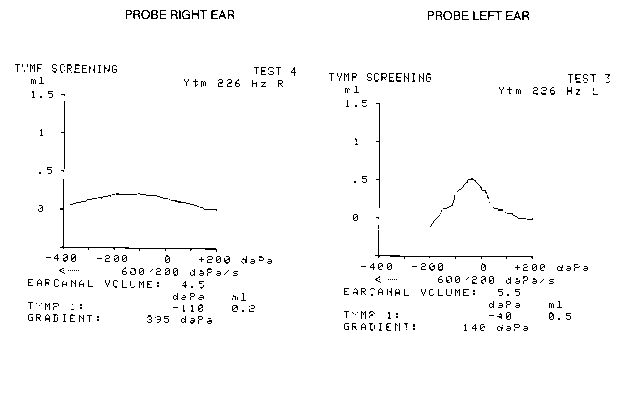
CASE STUDY: Modified Radical MastoidectomyRoy F. Sullivan, Ph.D. (posted 03/14/1996; revised 03/06/1999) This case is a 69 year old female patient with a history of chronic, bilateral middle ear disease beginning in childhood and continuing through middle age. Bilateral modified radical mastoidectomies were performed with removal of extensive cholesteatomae in 1989 (AD) and 1990 (AS). Under regular ENT care for mastoid cavity hygiene, both ears have been effusion-free for the last few years. The patient has used a Class II (vented) full concha hearing aid on the right ear for four years. The aid had been lost at time of evaluation and was subsequently replaced. The inline images below (23k) show the modified radical mastoidectomy cavities of both right and left ears. Note the exposed mastoid air cells lined with mucous membrane. Tympanic membranes are intact in both ears. Aural acoustic immittance (6k) shows a tympanogram peaked near ambient air pressure (Type A) on AS and a shallow, rounded tympanogram (Type B) centering on -180 daPa. Note the enlarged equivalent volume (PVT) values characteristic of the modifed radical and radical mastoidectomies; 4.5ml AD and 5.5ml, AS. Pure tone audiometry (23k) belies the extensive surgical revision of EAC and mastoid anatomy; the middle ear anatomy essentially spared in the modified radical procedure. 
|