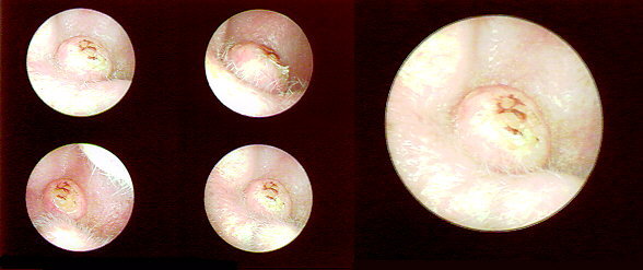
Thumbnail and CU view of Keratoacanthoma
The patient is a 61 year old man with a 6 week history of a painful, progressively expanding mass in his right ear. Its growth had stabilized over the last 2 weeks. Physical exam revealed a 1 cm smooth, raised mass which was centrally ulcerated and umbilicated located posteriorly and inferiorly in the concha. It measured 11mm x 6mm x 4mm.
Preoperative diagnosis was a probable keratoacanthoma. The lesion was excised and the defect was repaired with a full thickness skin graft. Pathological examination revealed it to be an irritated keratoacanthoma with associated inflammatory cells. The patient will be followed since this type of lesion can be difficult to differentiate from a well differentiated squamous cell carcinoma even with careful microscopic analysis.