Mural Cholesteatoma 12/97
The patient is a 13 year old female who was being seen by the ENT Department for follow up care since undergoing a canal up [intact wall] mastoidectomy and tympanoplasty at age 11.5 yrs. During her initial surgery a massive amount of cholesteatomatous material and granulation tissue was removed from the middle ear. The long process of the incus had been eroded by the cholesteatoma but was used to reossicularize the chain following its reshaping and repositioning between the neck of the malleus and the capitulum of the stapes. The defect in the TM was grafted and a collar tube was inserted.
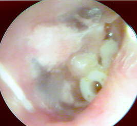
At her second 6 month check-up it was noted that a small mural cholesteatoma had developed anterior to the handle of the malleus and posterior to the tube. A VO picture of the mural cholesteatoma was taken in December 1997.
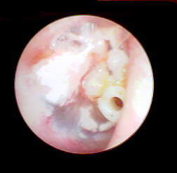
Her surgery was delayed but was rescheduled for May 1998. A second VO picture documents no change in the size of the mural cholesteatoma.
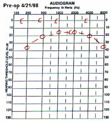
A preop audiogram was also obtained which was unchanged from the post-op audiogram of a year earlier.
During surgery, the small white mass was dissected away from the TM and a plane was established between the surface of the rotated incus and this mass. The mural cholesteatoma was also attached to the undersurface of the outer phalange of the tympanostomy tube. The tube was carefully elevated and removed along with the mural cholesteatoma. The middle ear was further explored after a whitish mass was seen through the thin posterosuperior quadrant of the TM. A middle ear cholesteatoma was removed. It had not yet displaced the ossicular reconstruction performed during the initial surgery and a new collar tube was inserted.
The pathology report described a 3mm mural choleateatoma consisting of partially cystic keratin debris. The middle ear cholesteatoma was 4mm and consisted of proliferative squamous lining.